Will Imaging Guide Future Depression Care?
Abstract
Rather than assess differences in volume or activity of targeted brain regions, exploring how the brain’s network is connected is emerging as a key research directive in neuroimaging.
In the search for biomarkers that can help predict how patients with depression will respond to medication, many researchers believe neuroimaging is the path that might offer the most promise.

So far, though, neuroimaging research looking for brain biomarkers that predict treatment response has not yielded much fruit. Studies have looked at both structural differences (such as hippocampal volume) and changes in brain activity, but results have been mixed.
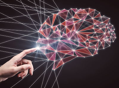
Conor Liston, M.D., Ph.D., an assistant professor of neuroscience and psychiatry at Weill Cornell Medical College, says that the inconsistent findings reflect the fact that a brain is not a set of discrete components doing their own thing. Rather, the brain is an interconnected circuit. “The question then becomes how do these components work together as a network?”
By using imaging tools, particularly functional magnetic resonance imaging (fMRI), researchers can assess functional connectivity—the strength and frequency of communication between areas of the brain involved in similar functions.
Scientists believe that there may be subtle, but identifiable, differences in the brain connection patterns among people with depression. Such differences may explain why depression symptoms differ significantly from patient to patient, or why patients who appear to have similar symptom profiles respond differently to medication.
Liston recently led an fMRI-based study that identified four distinct connectivity subtypes among depression patients; each subtype was found to be associated with a slightly different clinical profile and different response rate to transcranial magnetic stimulation (Psychiatric News, February 3). With more imaging samples and refined tools to analyze brain networks, Liston said that he believes that many more depression subtypes will emerge.
Boadie Dunlop, M.D., an associate professor of psychiatry and behavioral sciences at Emory University and director of its Mood Disorders Program, has also been working in this field and recently published some findings that show connectivity patterns in the subcallosal cingulate cortex can distinguish people with depression who respond to treatments from those who do not (Psychiatric News, May 19).
What was especially encouraging about Dunlop’s study is that the fMRI scans could identify the potential success or failure of both antidepressants and cognitive-behavioral therapy; previous imaging work had focused on only one intervention.
“In this [scenario], we can move a lot more people forward in their treatment since we can guide them toward medication or psychotherapy depending on their connectivity scores,” Dunlop said.
Still, Dunlop noted that more research is needed before fMRI can be used to guide treatment recommendations. Some patients in his study had scores that fell into a “gray zone” where a prediction could not be made. For those patients with clearer outcomes, the accuracy of the scans in determining treatment response or failure averaged around 78 percent.
That is a decent accuracy rating, but perhaps not good enough given the time and cost of fMRI scans, said Scott Langenecker, Ph.D., an associate professor of psychiatry and psychology at the University of Illinois at Chicago (UIC) who also directs UIC’s Cognitive Neuroscience Center.
“It turns out that there are many imaging-based models that work pretty well at finding average changes and predicting treatment response at a group level,” he said. “But none so far have been accurate enough to be clinically viable for individual patients. Is there a way we can get more information from these networks to get that extra level of precision?”
Langenecker and colleagues are aiming to answer that question by adding synchronicity (how efficiently these brain networks are synced when communicating) to their connectivity analyses in addition to the location and relative strength of functional connections. They do this by giving subjects cognitive tasks and observing the signal coordination between the error detection network and the interference processing network (which focuses the brain in the face of multiple tasks) when a mistake is made.
They found that people with depression who had stronger activity in either network during these tasks tended to respond less to antidepressants. People with strong activity and signals that were out of sync had even lower antidepressant response rates. When including synchronicity signatures in the prediction models, Langenecker could reach about 90 percent accuracy in identifying treatment response.
“It’s very close, but not quite there yet,” Langenecker said of the clinical potential of his model, though he added that he can’t guarantee a similar accuracy if he were to scan another set of participants, which he hopes to do. “But at this point the work is still more about discovery than application; we need to find out which networks are most important in depression and other mental illnesses.”
Besides, Langenecker said he thinks that the costs of fMRI are still too prohibitive at this point to guide treatments. In the future, as MRI devices become less expensive and more transportable, he expressed optimism that neuroimaging could become a more routine component of care.
Dunlop is a little less optimistic about how widely fMRI scanning can be used. “I think the real-time imaging required will remain complex in terms of processing time, and that will prevent it from becoming a routine procedure,” he said. “Ultimately, our goal should be to identify a non-imaging surrogate, maybe something in the blood for example, that reflects what we see in the brain.”
As with Langenecker, Dunlop said he agrees that investigators need to know more about these functional connectivity patterns before having any discussions about their role in developing treatments.
His team is pursuing several projects, but one of significant interest is seeing if an individual’s connections change during and after treatment. “This will address whether the connectivity patterns we see indicate a person’s trait or just a person’s state,” he said. ■


