Poor Intra-Brain Synchrony May Underlie Autism
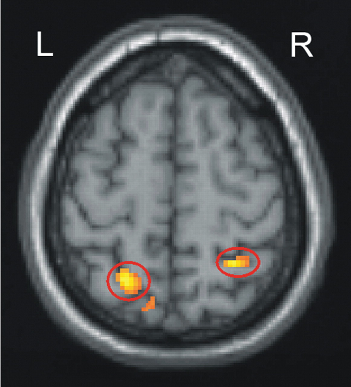
Study participants with autism showed greater activation in areas near the intraparietal sulcus (circled regions) than normal control participants during the processing of low-imagery sentences. The areas near the intraparietal sulcus are associated with visual imagery.
Source: Marcel Just, Ph.D., Brain advance-access Internet site.
A decade ago a woman with autism wrote: “I think in pictures. Words are like a second language to me. I translate both spoken and written words into full-color movies, complete with sound, which run like a VCR tape in my head.”
Thanks to neuroimaging, scientists are starting to confirm what this woman and others with autism have claimed—that they tend to think visually.
Last year, for example, an fMRI study headed by Nancy Minshew, M.D., a professor of psychiatry and neurology at the University of Pittsburgh, and Marcel Just, Ph.D., a professor of psychology at Carnegie Mellon University, showed that when individuals with autism attempt to recall letters of the alphabet, their brains process the stimuli differently from the brains of nonautistic individuals. The former rely more on the parietal regions, which are involved in visuospatial processing, whereas the latter depend more on the left prefrontal cortex, which is involved in language processing (Psychiatric News, February 4, 2005).
Now a new fMRI study conducted by the same group, and published on the Brain advance-access Internet site on July 10, reveals that when persons with autism attempt to comprehend sentences, they rally visual centers in their brains to do so even if the sentences do not contain visual images.
For example, one of the low-imagery sentences used in the experiment was“ Addition, subtraction, and multiplication are all math skills.” As the control group comprehended this sentence, their left prefrontal cortex was activated. But as the autism group comprehended this sentence, not only their left prefrontal cortex, but their parietal regions were switched on as well.
Moreover, as the autistic prefrontal cortex and parietal regions process language or engage in other cerebral challenges, they may be out of step with each other, another investigation has found. It was conducted by the same research team and was published on the Cerebral Cortex advance access Internet site on June 13.
In this inquiry, fMRI was used to examine the brains of both autism subjects and control subjects as they performed the Tower of London test. This task involves moving three balls into a specified arrangement. It requires planning and goal-management ability, using both the prefrontal cortex and the parietal regions. In the control group, the prefrontal cortex and parietal regions tended to function in synchrony—that is, increasing or decreasing their activity at the same time—whereas in the autism group, these two brain areas were less likely to do so.
Out of Sync Due to Faulty Connections
This lack of synchronization between the prefrontal and parietal regions, in turn, implies that it may be due to faulty white matter fiber connections between them. The Cerebral Cortex study supports this supposition. The largest white matter fiber tract in the brain is the corpus callosum, which allows communication between the two sides of the brain. One part of the corpus callosum is the genu. The genu was found to be smaller in the autism group than in the control one, and the smaller the genu in the autism subjects, the more out of sync their frontal and parietal regions were.
Of all their new findings, Just considers this to be the most important, he told Psychiatric News. “The reason that this finding is important—and rare—is that it makes a crucial link between [the synchronization of activity between different brain areas] and the anatomical white matter brain structure—corpus callosum—that carries the communication between the areas. It is a crucial link between brain activation and brain tissue in autism.”
Autism: Lack of Brain Communication?
Indeed, when all of these findings are considered together, they make Minshew, Just, and their group wonder whether a reduced white fiber connectivity might underlie the neurological, psychological, and behavioral symptoms that constitute the syndrome of autism. And if autism is truly due to a failure of various brain regions to communicate with each other, it“ may one day provide the basis for improved treatments for autism that stimulate communication between brain areas,” Duane Alexander, M.D., envisions. He is director of the National Institute of Child Health and Human Development. That institute, as well as the National Institute on Deafness and Other Communication Disorders, funded the two studies.
Meanwhile, Minshew told Psychiatric News that these study results help explain why individuals with autism can be so bright in specialized ways, yet have trouble with problem-solving and understanding the gist of things—they have the local brain connections to perform the former, but not the widespread brain connections to carry out the latter.
“So in dealing with patients with autism,” Minshew advised,“ say whatever you have to say as simply as possible. Don't give a lot of examples and expect them to extract the concept [because the circuits that handle these functions may not be working properly]. Also, don't expect them to generalize from one experience to the next [for the same reason]. Use facts, details, and their favorite interest to motivate them.”
An abstract of “Sentence Comprehension in Autism: Thinking in Pictures With Decreased Functional Connectivity” is posted at<http://brain.oxfordjournals.org/cgi/content/abstract/awl164v1>. An abstract of “Functional and Anatomical Cortical Underconnectivity in Autism: Evidence From an fMRI Study of an Executive Function Task and Corpus Callosum Morphometry” is posted at<http://cercor.oxfordjournals.org/cgi/content/abstract/bhl006v1>.▪