Infants’ Eyes May Reveal Clue to Autism Risk
Abstract
Eye movements and visual attention are measurably delayed in infants who are later diagnosed with ASD.
Abnormal oculomotor functioning in infants may portend a diagnosis of autism spectrum disorder (ASD). Researchers participating in the Infant Brain Imaging Study (IBIS) reported in the March 20 AJP in Advance that seven-month-old infants who are eventually diagnosed with ASD take a split second longer to shift their gaze during a task measuring eye movement and visual attention than do typically developing infants of the same age.
The gaze delay lasts only 25 to 50 milliseconds on average, too brief to be detected in normal social interactions with an infant. But lead author Jed Elison, Ph.D., of the University of North Carolina at Chapel Hill and the California Institute of Technology and colleagues report that the delay is measurable and could possibly be accounted for by differences in the structure and organization of actively developing neurological circuits of a child’s brain.

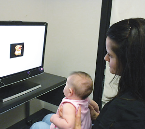
Infants were shown images on a screen while their eye movements were recorded and measured.
Their data were collected from 97 infants, of whom 16 were considered to have high familial ASD risk due to having an older sibling already diagnosed with ASD and who were later diagnosed as having ASD themselves. Another 40 were at high familial risk but did not later meet ASD criteria, and 41 were low-risk infants. All of the infants were enrolled at approximately 6 months of age and underwent a comprehensive behavioral assessment and brain imaging protocol at the ages of 6, 12, and 24 months. The data used in the analysis included behavioral, imaging, and eye-tracking data from the initial six-month visit and clinical assessment data from the 24-month visit.
Imaging methods assessed radial diffusivity (an index of white matter organization) in fiber tracts that included corticospinal pathways and the splenium and genu of the corpus callosum. The children were assessed at the University of North Carolina at Chapel Hill and Children’s Hospital of Philadelphia. The study was conducted in the context of an ongoing Autism Center of Excellence Network study, which was prospectively investigating longitudinal brain and behavioral trajectories in high-familial-risk infant siblings of children diagnosed with ASD and low-risk comparison infants.
Visual orienting delays were longer in 7-month-old infants who later showed ASD symptoms, compared with both high-risk negative infants and low-risk infants. The research also indicated that the size of the splenium was associated with the speed with which the low-risk infants were able to shift their gaze, but in infants later found to have ASD there was no correlation between splenium size and speed of gaze shift.
The researchers theorized that the differences in gaze shifting between the two groups may not be due directly to differences in the splenium between the groups, but to differences in the brain circuit that connects the splenium to visual areas of the brain.
Could oculomotor assessment of infants enhance early identification of those who develop ASD? Elison and his colleagues hope for more than that: “The present behavioral findings not only complement and extend recent data suggesting early functional and structural brain abnormalities in infants who go on to develop ASD,” they said, “they also have the potential to enhance early identification of individuals with ASD and to inform targeted interventions developed for the ASD prodrome.” They noted that one recent study successfully altered visual orienting performance in 11-month-old infants using several gaze-contingent attention training procedures administered during five visits over 15 days. “Additional research is needed to determine the long-term effects of modifying visual orienting patterns in infancy, specifically in relation to how training attention at specific time intervals during infancy might alter social-cognitive development,” they concluded.
The research is part of the ongoing Infant Brain Imaging Study, which is supported through the National Institute of Child Health and Development’s Autism Centers of Excellence Program, Autism Speaks, and the Simons Foundation. ■
An abstract of “White Matter Microstructure and Atypical Visual Orienting in 7-Month-Olds at Risk for Autism” is posted at http://ajp.psychiatryonline.org/article.aspx?articleid=1669751. Information about the Infant Brain Imaging Study (IBIS) Network consortium is posted at http://www.ibisnetwork.org/.


