Neuroscience 2014 Showcases Advances in Mental Health Research
Abstract
Knowledge about structure, development, health, and diseases of the brain and nervous system is advancing rapidly, according to research by some of the world’s leading neuroscientists.

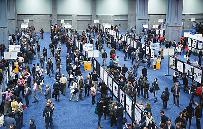
More than 30,000 scientists converged in Washington, D.C., in November for Neuroscience 2014, the largest conference devoted to fundamental and translational research on the brain and nervous system, offering some of the first glimpses of tools and discoveries that may lead to future clinical breakthroughs.
What follows are summaries of just a few of the research studies advancing knowledge of mental health.
Cerebellar Volume Tied to Intellectual Disability, but Not Austism
The cerebellum, a brain region involved in motor control, has been thought to play a significant role in autism spectrum disorder (ASD), but research so far has been conflicting. A new brain imaging study—one of the largest related to ASD to date—suggests that the discrepancies arise because cerebellar abnormalities are associated with intellectual disability, but not ASD as a whole.
Researchers at the Hospital for Sick Children in Toronto examined magnetic resonance images using a new cutting-edge program that enabled them to measure the volume of the cerebellum’s subregions. Their analysis of 333 people with ASD and 376 controls found that cerebellar volumes were not significantly different between the two groups. However, both groups did show an association between cerebellar volumes and IQ.
In addition, in two subregions—the left inferior posterior and left anterior lobes—tissue volume predicted IQ more poorly in ASD subjects than in healthy subjects, suggesting a target for future studies.
Park MM, Lerch JP, et al. Cerebellar involvement in autism spectrum disorders and cognitive ability. Center for Addiction and Mental Health and Program in Neuroscience and Mental Health, Hospital for Sick Children, Toronto
Childhood Maltreatment May Alter Circuits in Prefrontal Cortex
Adolescence is a critical period in brain development, especially for areas that regulate emotions and impulse control; concurrently, adolescence is also a time when disorders like depression or addictive behaviors begin to manifest.
New work from Yale University suggests that childhood maltreatment affects how parts of the brain develop in later years. Forty-four teens with a history of childhood abuse or neglect received brain scans and behavioral assessments at around ages 15 and 18.
The scans revealed that maltreatment lowered the structural integrity of white matter in parts of the prefrontal cortex, as well as some brain regions connected to the cortex. The patterns were different in teens reporting physical versus emotional abuse, and in the latter instance, differences were also observed in how the brains of men and women responded to emotional maltreatment.
These data offer a physiological mechanism supporting findings that males who have been mistreated tend to develop externalizing problems like drug use, while mistreated females develop internalizing problems like depression.
Cox ET, Wang F, et al. Child maltreatment and corticostriatolimbic development in adolescence: Effects of maltreatment subtype and gender. Yale University
Visual Test Can Distinguish People With ADHD Versus Bipolar Disorder
An eye-movement task incorporating emotional faces could differentiate bipolar disorder (BD) from attention-deficit/hyperactivity disorder (ADHD). While BD and ADHD are distinct types of mental disorders, they have several cognitive and emotional symptoms in common. This suggests that eye-movement tests could provide some nonsubjective information for distinguishing or diagnosing psychiatric illnesses.
The study, carried out by a group at Queen’s University in Canada, used a modified version of the antisaccade task, in which subjects try to look away from a visual cue that had emotional faces in the background. Both subjects with ADHD and with BD had slower reaction times and made more errors then controls, and they each showed some emotion-specific differences. Fearful faces increased direction errors and saccadic reaction times for ADHD subjects, while neutral faces had the same effect on BD subjects.
These findings suggest that combining cognitive and emotional tasks can help identify nuanced deficits in how the brains of people with specific mental disorders process information. The researchers hope to expand their work to see how conditions that are more closely related, for example BD and major depressive disorder, respond to these tests.
Soncin S, Brien D., et al. Saccidic eye movement tasks help differentiate ADHD and bipolar disorder. Center for Neuroscience Studies, Queen’s University, Kingston, Ontario
New Imaging Technique Identifies Brain Cells Involved in Eating
Using an advanced bioimaging technique, a team at the University of North Carolina has uncovered specific brain cells that contribute to hunger and feeding. This work opens up new prospects for understanding the neurological basis of eating disorders such as anorexia or bulimia.
While it is known that the brain’s lateral hippocampus is involved in eating, researchers do not know which cells regulate the specific aspects of eating behavior. To try and identify these cells, the researchers developed a mouse model with engineered neurons that would light up whenever they triggered a calcium wave, signaling that a neuron is firing.
The researchers then implanted a microendoscope near these neurons so they could see which neurons glowed as the mice roamed around and ate. With this approach, they could distinguish which cells were active when the mice were eating compared with when they were trying to gain access to food.
By gaining a better sense of how the circuitry operates when feeding behaviors are normal, the researchers can examine what goes wrong in the brain cells when an eating disorder is present.
Stuber GD, Jennings JH, et al. Deep brain calcium imaging of distinct lateral hypothalamic neuronal ensembles reveals a precise neural signature for motivation. Departments of Psychiatry, Radiology, Bioengineering, Univ. of North Carolina at Chapel Hill and Inscopix, Palo Alto, Calif.
Diet Influences Long-Term Behavior After Concussion
Some children experience a concussion or other physical brain injury as they mature; in most cases they recover completely, but a significant proportion experience lingering, and often permanent, symptoms related to memory, thinking, and behavior. The question becomes how to identify such children and mitigate long-term effects.
A new study in mice from researchers at the universities of Lethbridge and Calgary in Alberta, Canada, points to a healthy diet as one way to reduce the risks of persistent neurological problems. Pregnant female mice were fed either a high-fat and high-sugar diet, a low-calorie diet, or a standard diet during pregnancy, and their offspring were given the same diet. At four weeks (equivalent to about eight to 11 years in a human), half of the offspring received a mild concussion.
Following the concussion, animals on the high-fat and high-sugar diet had persistent problems with balance, anxiety, and executive functioning, while the mice on the low-calorie diet showed almost no deficits on those same tasks.
This study reinforces other work showing a connection between proper nutrition and neurological health, though it is new in the context of concussions. The researchers next want to test if switching to a low-calorie diet after a concussion can also improve brain recovery. ■
Mychasiuk RM, Hehar H, et al. Caloric intake affects behavioural outcomes following pediatric concussion in rats. University of Calgary and the University of Calgary School of Medicine.


