Journal Digest
Altered Brain Activity Found to Impact Behavioral Control

Adolescence is a period of heightened exploration and novelty seeking, which can increase the risk of potentially dangerous behaviors.
A study appearing in Current Biology now provides some of the first causal evidence linking risk-taking behaviors with an imbalance in brain activity in the prefrontal cortex (PFC)—which is involved in cognitive control and inhibition—and the nucleus accumbens (NAC)—which plays a key role in reward seeking.
For the study, a team of researchers at Dartmouth College used an approach known as chemogenetics (drugs are used to turn genes in a specific region off or on for a limited time) to simultaneously decrease activity in the orbitofrontal cortex (OFC) and increase activity in the NAC while adult rats learned a task.
They tested the effects of this imbalance in an inhibition task, in which rats would receive food following a tone but no food if there was a light then a tone (thus testing the ability of the animals to withhold their desire for food in appropriate situations).
Rats who had reduced OFC activity and heightened NAC activity had a harder time learning to withhold an inappropriate response—an effect that was greater than that of either manipulation alone. In fact, adult rats with both brain regions altered took about as long to learn as seen in normal adolescent rats.
“These findings provide direct evidence that simultaneous underactivity in OFC and overactivity in NAC can negatively impact behavioral control and provide insight into the neural systems that underlie inhibitory learning,” the authors wrote.
Meyer H, Bucci D. Imbalanced Activity in the Orbitofrontal Cortex and Nucleus Accumbens Impairs Behavioral Inhibition. Curr Biol. September 20, 2016. pii: S0960-9822(16)30978-2.
Targeted ApoE4 Reduction in Brain Does Not Inhibit Learning, Memory

Therapies that target apolipoprotein E4 (ApoE4)—a known risk factor for the development of Alzheimer’s disease—might prove useful in delaying or preventing disease onset, but it’s not known how critical the protein is for normal brain function. A study in the Journal of Neuroscience suggests that removing most of ApoE in the brain may have little impact on learning and memory in mice.
Previous studies show ApoE knock-out mice have synaptic loss and cognitive dysfunction; however, these findings are complicated by the fact that ApoE knock-out mice have highly elevated plasma lipid levels, which may independently affect brain function.
To evaluate the effects of ApoE loss in the context of a normal lipid profile, researchers at the University of Texas Southwestern Medical Center developed a mouse model that expresses ApoE normally in peripheral tissues, but has severely reduced ApoE in the brain.
While these mice displayed abnormalities in synaptic connections, they showed no impairments in learning or memory, and had the same ratio of AMPA/NMDA receptors as control mice. The mice with reduced ApoE in the brain also retained normal lipid profiles, unlike mice with a full ApoE4 knockout which develop severe atherosclerosis.
“These findings suggest that plasma lipid levels can influence cognition and synaptic function independent of ApoE expression in the brain,” the authors wrote.
Lane-Donovan C, Wong W, Durakoglugil M, et al. Genetic Restoration of Plasma ApoE Improves Cognition and Partially Restores Synaptic Defects in ApoE-Deficient Mice. J Neurosci. September 28, 2016; 36(39):10141-50.
Researchers Identify Schizophrenia Risk Genes That Alter Neurodevelopment

Recent genomic efforts have uncovered over 100 gene variants associated with schizophrenia risk, yet how these variants exert their influence remains uncertain.
A study published in Nature Neuroscience has now identified five genes from this extensive list that could have significant biological impact.
Researchers sequenced RNA (an indicator of gene expression) from the dorsolateral prefrontal cortex from the postmortem brain tissue from people with schizophrenia and controls.
They found that of the 108 variants previously linked to schizophrenia, 20 appeared to regulate gene expression, and five of these variants could be pinpointed to a specific gene (FURIN, TSNARE1, CNTN4, CLCN3, or SNAP91).
Altering the expression of FURIN, TSNARE1, and CNTN4 in zebrafish was associated with changes in neurodevelopment. The researchers also found that depleting FURIN in human neural progenitor cells decreased the migration of these cells in culture—hinting at a possible mechanism by which schizophrenia might disrupt the developing brain.
“As we move from gene discovery—which for risk genes can be done in any tissue—to the human brain, we are able to explore changes in the molecular machinery of the cell, such as gene expression and epigenetic mechanisms, that are impacted by these genetic risk factors,” Thomas Lehner, Ph.D., director of the National Institute of Mental Health Office of Genomics Research Coordination, said in a press statement.
Fromer M, Roussos P, Sieberts S, et al. Gene Expression Elucidates Functional Impact of Polygenic Risk for Schizophrenia. Nat Neurosci. September 26, 2016. [Epub ahead of print]
Childhood Stress May Accelerate Telomere Shortening

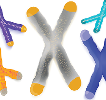
The shortening of telomeres, the protective caps on the ends of chromosomes, occurs as cells age and is linked with many diseases, including neuropsychiatric disorders. While stressful events are believed to accelerate cellular aging, few studies have explored the relationship between telomere length and accumulated stress.
A study published in the Proceedings of the National Academy of Sciences suggests adverse experiences in childhood may increase the likelihood of having short telomeres later in life.
For the study, researchers compared the telomere lengths of 4,598 men and women (50 and up) who were enrolled in the U.S. Health and Retirement Study. As part of this study participants had self-reported various financial, social, and traumatic events during childhood and adulthood.
The findings revealed that accumulated adverse experiences in childhood significantly predicted an increased likelihood of having short telomeres later in life. The occurrence of each additional childhood event predicted an 11 percent increased odds of having short telomeres.
Follow-up analysis that categorized the stressors showed that the increased odds were principally associated with social or traumatic stress during childhood and not financial stress.
“Not all children who experience adversity are at similar risk… Researchers have identified genetic, psychosocial, and behavioral factors that may mitigate or potentiate vulnerability to adverse experiences, and understanding their complex interplay will allow us to optimize policy initiatives for interventions for families … that target those most at risk for excessive early morbidity and mortality,” the authors wrote. ■
Puterman E, Gemmill A, Karasek D, et al. Lifespan Adversity and Later Adulthood Telo-mere Length in the Nationally Representative US Health and Retirement Study. Proc Natl Acad Sci USA. October 3, 2016 [Epub ahead of print]


