Journal Digest
Binge Drinking Induces Sex-Specific Gene Expression Changes

A genetic study in mice suggests that binge drinking has different biological effects in the brains of males and females. These findings were published in Frontiers in Genetics.
Researchers at Oregon Health and Science University compared gene expression in mice given access to ethanol with mice given access to water only. To recreate binge-drinking conditions during the study, the animals received limited access to fluids. Every three days, the animals were given 30 minutes of unlimited access to water infused with ethanol or water only. A total of nine males and nine females were included in each group.
The researchers specifically looked at the expression of 384 genes in the nucleus accumbens—a brain region linked with addiction. They found that the expression of 106 genes was altered by binge drinking, but the expression of only 14 of these genes was altered in both males and females; 56 of the 106 genes were altered by binge drinking in females only and 36 genes in males only. Of the 14 genes altered in both sexes, only four had the gene expression change in the same direction (more expression or less expression) in response to binge drinking.
The gene expression profiles suggested that hormone signaling and immune function were altered by binge drinking in female mice, whereas neurotransmitter metabolism was primarily affected by binge drinking in male mice.
“[A]n increased understanding of sexually dimorphic molecular pathways influenced by binge drinking and chronic intoxication leading to dependence may identify novel treatment options for males and females,” the researchers wrote.
Finn DA, Hashimoto JG, Cozzoli DK, et al. Binge Ethanol Drinking Produces Sexually Divergent and Distinct Changes in Nucleus Accumbens Signaling Cascades and Pathways in Adult C57BL/6J Mice. Front Genet. September 10, 2018. [Epub ahead of print]
Nicotine Therapy May Improve Symptoms of Late-Life Depression

A pilot study by researchers at Vanderbilt University Medical Center suggests that adjunctive transdermal nicotine might improve mood and cognition in older patients with depression.
Late-life depression is characterized both by mood symptoms and cognitive deficits, the latter of which are believed to hinder the effectiveness of antidepressants and increase the risk of depression relapse. Nicotine has been shown to increase activity of brain networks involved in cognitive control.
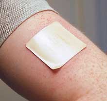
Fifteen adults aged 60 and older with major depressive disorder and cognitive impairment were enrolled in the study. All participants were nonsmokers and were on stable doses of antidepressants. Nicotine patches were applied daily over 12 weeks and titrated up to a maximum dose of 21 mg nicotine a day. Patients were evaluated using cognitive tests and mood scales every three weeks.
At 12 weeks, 13 of the 15 participants achieved a response to their antidepressant (defined as at least a 50 percent reduction in Montgomery-Åsberg Depression Rating Scale [MADRS] scores). Eight participants achieved depression remission (MADRS score of 8 or lower). The participants also showed improvements in some tests involving working memory, episodic memory, and emotional processing, though not in attention.
This study was published in the Journal of Clinical Psychiatry.
Gandelman JA, Kang H, Antal A, et al. Transdermal Nicotine for the Treatment of Mood and Cognitive Symptoms in Nonsmokers With Late-Life Depression. J Clin Psychiatry. 2018; 79(5) pii: 18m12137.
Children With ASD Express Positive Emotions Similar to Non-ASD Toddlers

There is a prevailing notion that children with autism spectrum disorder (ASD) exhibit intense negative emotions and blunted positive emotions, yet little research has explored emotional expression in this group.
A study by a research team at the Yale Child Study Center now suggests toddlers with ASD have muted responses to threats, while their ability to express positive emotions is similar to normal toddlers.
The study included 43 toddlers with ASD, 16 with developmental delay (DD), and 40 with typical development (TD). The investigators used standard cues to induce anger, fear, and joy in the toddlers while recording facial and vocal responses.
The researchers observed that toddlers with ASD exhibited less intense fear compared with toddlers with DD and TD and more intense anger than toddlers with DD, but not those with TD; children in all three groups were equally expressive of joy. The intensity of fear and anger in ASD children was not related to symptom severity, but joy intensity was, with greater symptom severity equated to less intense joy.
“The results reveal a complex and surprising emotional landscape of very young children with ASD, a landscape that is inconsistent with the negative emotionality bias hypothesis and largely independent of autism symptom severity,” the researchers wrote.
The findings were published in the Journal of the American Academy of Child and Adolescent Psychiatry.
Macari S, DiNicola L, Kane-Grade F, et al. Emotional Expressiveness in Toddlers With Autism Spectrum Disorder. J Am Acad Child Adolesc Psychiatry. August 29, 2018. [Epub ahead of print]
Calculator Predicts Dementia Risk Rates for ApoE4 Carriers

People with one or two copies of the E4 variant of the apolipoprotein E gene (ApoE4) have a higher risk of Alzheimer’s disease.
In a study appearing in the Canadian Medical Association Journal, researchers at Copenhagen University Hospital used data from two large Danish health studies to quantify 10-year dementia risk based on ApoE4 status. The combined studies included 104,537 individuals, of whom 2,160 developed dementia and 7,520 developed cerebrovascular disease. All the participants underwent genetic tests to identify their ApoE status (whether they have the E2, E3, or E4 alleles).
The researchers found that, as expected, people with E4/E4 had the highest 10-year Alzheimer’s risk, followed by those with the E4/E3, E4/E2, E3/E3, E3/E2, and E2/E2 alleles. The absolute 10-year Alzheimer’s risk for women with the E4/E4 genotype was 7 percent at ages 60 to 69, 16 percent at ages 70 to 79, and 24 percent at 80 years or older; for men, the corresponding risks at these ages were 6 percent, 12 percent, and 19 percent, respectively.
The 10-year risk for any dementia in E4/E4 carriers was 10 percent, 22 percent, and 38 percent for women 60 to 69, 70 to 79, and 80 years or older, respectively. The 10-year risk for any dementia in men who were E4/E4 carriers was 8 percent, 19 percent, and 33 percent for men 60 to 69, 70 to 79, and 80 years or older, respectively.
“Such estimates have the potential to facilitate the identification of high-risk individuals for targeted preventive interventions,” the researchers wrote.
Rasmussen KL, Tybjærg-Hansen A, Nordestgaard BG, Frikke-Schmidt R. Absolute 10-Year Risk of Dementia by Age, Sex, and APOE Genotype: A Population-Based Cohort Study. CMAJ. 2018; 190(35): E1033-E1041.
Antipsychotic-Associated Hyperprolactinemia Increases Health Care Costs

A health insurance analysis appearing in the Journal of Medical Economics reports that antipsychotic-associated hyperprolactinemia is associated with substantial health care costs. Hyperprolactinemia involves having elevated levels of the hormone prolactin in the blood, which can result in sexual and fertility problems.
Researchers at Analysis Group Inc. in Montreal used data from a large U.S. insurance claims database to identify 499 patients (242 on commercial insurance and 257 on Medicaid) who developed hyperprolactinemia while taking antipsychotics. Next, they assessed the health care costs for these patients over the six months following their hyperprolactinemia diagnosis and compared them with the six-month costs of a matched set of patients taking antipsychotics but hyperprolactinemia-free.
Among commercially insured patients, hyperprolactinemia was associated with additional medical costs of $3,861 ($13,218 versus $9,357) and additional total health care costs of $5,732 ($20,081 versus $14,349). In Medicaid patients, the added medical and total health care costs of hyperprolactinemia were $9,246 ($20,859 versus $11,613) and $10,773 ($30,763 versus $19,990), respectively. Inpatient care costs were a significant contributor to these differences.
The researchers also compared the medications taken in the hyperprolactinemia and hyperprolactinemia-free cohorts. Based on some calculations, they estimated that using an antipsychotic considered “prolactin sparing,” such as quetiapine, could reduce the risk of hyperprolactinemia by 80 percent.
Cloutier M, Greene M, Touya M, et al. A Real-World Analysis of Healthcare Costs and Relative Risk of Hyperprolactinemia Associated With Antipsychotic Treatments in the United States. J Med Econ. September 6, 2018. [Epub ahead of print]


