Journal Digest
Early Life Stressors Speed Up Adolescent Brain Pruning

Stressful life events in early childhood accelerate the maturation of certain brain regions during adolescence, reports a study appearing in Scientific Reports.
These findings came from a nearly 20-year longitudinal study of children conducted by researchers at Radboud University in the Netherlands. The researchers compared brain maturation, defined as changes in gray matter volume, in 37 children who had MRI scans taken at ages 14 and 17.
During adolescence, neural connections in the brain undergo pruning, resulting in less gray matter.
Adolescents who experienced more negative events such as serious illness or parent divorce during early childhood (defined as up to age 5 years) displayed faster gray matter volume (GMV) reduction in multiple brain regions between ages 14 and 17. In contrast, negative life events between 14 to 17 were not associated with reductions in GMV.
The researchers also found that experiencing a negative social environment (the overall quality of home and school interactions) between 14 and 17 appeared to slow down brain maturation in the anterior cingulate cortex. They noted this has significant implications as the anterior cingulate is associated with impulse control and emotional regulation and a slower maturation increases the risk of antisocial behaviors.
Tyborowska A, Volman I, Niermann H et al. Early-life and Pubertal Stress Differentially Modulate Grey Matter Development in Human Adolescents. Sci Rep. 2018; 8(1): 9201.
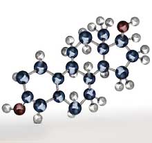
Estradiol May Worsen Anxiety Following Methamphetamine Withdrawal

A study published in Neuroscience Letters provides an explanation for why women may have more difficulty than men with methamphetamine withdrawal. The study, conducted by researchers at Dickinson College, suggests that the sex hormone estradiol worsens anxiety-like behaviors during drug withdrawal, which may increase the risk of relapse.
The researchers replaced ovaries in female mice with capsules that released either sesame oil or estradiol. The researchers let the mice play in an open place for three hours; then injected them with either low-, medium-, or high-dose methamphetamine; and placed the animals back in the open area for five more hours.
Both groups of mice behaved the same prior to the methamphetamine injections and during the first hour following the injection. Afterwards, as the drug started clearing their system, the mice with estradiol capsules who received high-dose methamphetamine were less active and spent more time on the perimeter of the open space compared with mice with sesame oil capsules. This effect was not seen for the low or medium doses of the drug.
The authors suggested that estradiol may exacerbate anxiety by activating the hypothalamic-pituitary-adrenal (HPA) axis in response to stress, though future studies are needed to confirm this.
Rauhut AS, Curran-Rauhut MA. 7 β-Estradiol Exacerbates Methamphetamine-Induced Anxiety-like Behavior in Female Mice. Neurosci Lett. 2018; 681:44-49.
Electrical Reward Signals May Predict Treatment Response

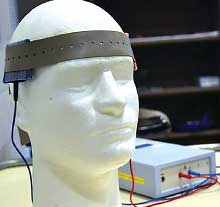
Depression and anxiety are associated with decreases in reward positivity, the electrical signal produced by neurons following a rewarding experience. A study published in Journal of Clinical Psychiatry has demonstrated that measuring reward positivity using electroencephalography (EEG) might be able to identify if a person with depression will respond better to an antidepressant or cognitive-behavioral therapy (CBT).
A team led by researchers at the University of Illinois at Chicago enrolled 63 volunteers with a diagnosis of anxiety or depression and 25 healthy controls. The participants first completed a short monetary-based computer game while wearing EEG caps to measure reward positivity. Afterwards, the people with depression or anxiety were randomized to receive 12 weeks of selective serotonin reuptake inhibitor (SSRI) therapy or CBT, after which point all participants conducted a second round of monetary games.
At baseline, higher depressive symptom scores were associated with lower reward positivity. After 12 weeks, there was a correlation between the degree in symptom improvement and improvement in reward positivity signals, regardless of treatment. The researchers also found that participants with a blunted positivity response before starting treatment experienced a greater reduction in depressive symptoms following treatment with SSRIs, but not CBT.
Burkhouse KL, Gorka SM, Klumpp H, et al. Neural Responsiveness to Reward as an Index of Depressive Symptom Change Following Cognitive-Behavioral Therapy and SSRI Treatment. J Clin Psychiatry. 2018; 79(4): 17m11836.
Transcranial Direct-Current Stimulation May Reduce Violent Impulses

A study in the Journal of Neuroscience suggests that transcranial direct-current stimulation (tDCS) to the prefrontal cortex may reduce the desire to commit violent acts.
Investigators at the University of Pennsylvania and Singapore’s Nanyang Technological University conducted a randomized, controlled trial of 81 healthy adults who received either 20 minutes of tDCS to the prefrontal cortex or a sham stimulation prior to participating in a behavioral task. The prefrontal cortex (specifically the dorsolateral prefrontal cortex) was chosen since it has been documented that antisocial individuals have deficits in this brain region that regulate complex thoughts and behavior.
After the stimulation, the participants were asked to rate the likelihood they might participate in two hypothetical scenarios, one related to physical assault and the other to sexual assault. They were also asked to rate how morally wrong each scenario was.
The participants receiving tDCS were 47 percent and 70 percent less likely than the sham group to report desires to participate in the physical and sexual assault, respectively. Based on the differences reported in the morality assessments, the researchers estimated 31 percent of the total effect of tDCS at reducing violent intentions was due to an increased perception of greater moral wrongfulness regarding the violent acts. ■
Choy O, Raine A, Hamilton RH. Stimulation of the Prefrontal Cortex Reduces Intentions to Commit Aggression: A Randomized, Double-Blind, Placebo-Controlled, Stratified, Parallel-Group Trial. J Neurosci. July 2, 2018. [Epub ahead of print]


