New Analysis Suggests There Is No Such Thing as Harmless Head Contact in Football
Abstract
Using innovative helmet-based sensors, researchers have pinpointed the midbrain as a key marker of early sports-related brain injury.
It may be cliché to term sports-related research as a “game changer,” but those words may accurately reflect the recent findings of investigators at the University of Rochester and Carnegie Mellon University (CMU) on the detrimental effects of sports-related head impacts.
With the help of specially designed football helmets with embedded sensors that monitor the speed and trajectory of incoming hits, this team found that just a single season of collegiate football results in damage to the brain’s white matter (nerve fibers that support the transmission of electrical signals throughout the brain)—even in the absence of a concussion or any outward symptoms of brain problems.

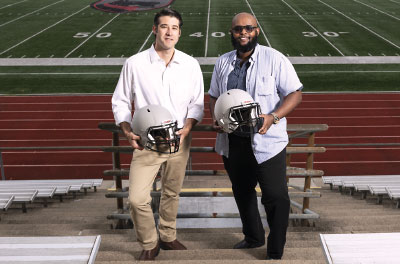
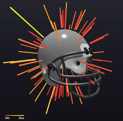
With the help of sensors that track real-time data on the hits a football player receives (see image below) over the course of a season, Bradford Mahon, Ph.D. (left), and Adnan Hirad, Ph.D., found that impacts that cause head rotations are more responsible for midbrain damage than head-on collisions.
What’s more, the researchers found that it’s not head-on collisions that appear to be the main culprits of damage to nerve fibers; rather, hits that result in head twisting seem to cause the most nerve damage. “Everyone interested in this area has focused on concussions, and that’s well and good,” noted lead investigator Adnan Hirad, Ph.D., an M.D./Ph.D. candidate at the University of Rochester who was a driving force behind the study. “But what about all these other repetitive hits sustained during games or practice. How bad are they [for the brain]?”
Based on the analysis of 38 University of Rochester football players who participated in a head impact study, these seemingly innocuous hits are anything but. MRI scans of these players taken before and after one football season revealed significant white matter damage in the midbrain, even though only two of the 38 participants sustained a concussion during the season. (The midbrain is the topmost section of the brainstem that controls sensory functions like vision, hearing, alertness, and the sleep/wake cycle.)
The degree of white matter damage correlated with both the number and force of hits sustained, especially the amount of rotational force a player sustained.
Senior investigator Bradford Mahon, Ph.D., an associate professor of psychology at CMU (and previously at Rochester), told Psychiatric News these findings raise several practical considerations.
For one, the detailed sensor readouts obtained in the study might lead to improved helmet designs that can reduce some of the potential damage. “Football helmets are heavy, and while that weight and thickness can reduce the effect of a linear impact, that added weight increases your susceptibility to a rotational shift,” he explained. “Developing lightweight helmets [using composite materials and force-absorbing foams or gels, for example] would help move the needle in regard to brain injury.

The sensors embedded in the helmet create a “spatial fingerprint” showing the direction and force of all sustained head hits. Over time, such information may help identify players at risk of clinical head trauma symptoms. See more at openbrainproject.org.
These helmet sensors might also serve as an objective way to measure an athlete’s neurological injury prior to the emergence of any clinical symptoms.
“My goal when starting the project was to find a good biomarker for [subtle] head injury,” Hirad said. “Is there an equivalent in the brain to the canary in the coal mine?”
Hiard focused on the midbrain because this region has multiple characteristics that make it a good location to assess early neurological damage. First, it is centrally positioned in the brain and susceptible to injury from multiple directions. The midbrain also regulates behaviors like eye movement, which is known to be disturbed after a concussion, so damage to this region likely contributes to the symptoms of head injury. Third, midbrain damage has been implicated in neurodegenerative diseases, which suggests that short-term physical damage can lead to chronic effects.
The strong connection between rotational impacts and white matter damage seems to validate Hirad’s choice of the midbrain as the proverbial canary. For further evidence, he and colleagues examined MRI scans from 29 other Rochester athletes from a variety of sports who sustained a concussion and observed that these athletes also had midbrain damage.
Mahon said he envisions a future where a football player can be monitored in real time using helmet sensors to assess potential brain damage without requiring an MRI scan. With such information, team doctors might be able to better identify when a player who has not sustained a concussion should be taken out of a game to prevent serious injury.
Study co-author Jeffrey Bazarian, M.D., a professor of emergency medicine at the University of Rochester, is also looking for blood-based biomarkers that correspond to impact-related neurological damage. (This would be useful in medical settings and for contact sports that don’t use helmets.) His latest research suggests that serum levels of the Alzheimer’s-related protein tau might be a good proxy for brain injury.
“Right now, the only criterion for playing is whether an athlete is showing any symptoms of a concussion,” Mahon said. “But that is the equivalent of sending someone with radiation exposure back into a nuclear environment just because the individual has no outward signs of radiation poisoning.”
Mahon noted that more data need to be collected to assess questions like how white matter damage accumulates on a week-to-week basis or if the midbrain can repair white matter damage following an extended contact-free offseason.
“But what we would really hope to do is monitor football players at different ages and assess them over multiple years,” he said. “With that, we might be able to get a glimpse of what a lifetime of football does to the brain, not just one season.”
As researchers come to understand football’s long-term effects on the brain, Mahon believes the discussion over football will have to tackle existential as well as practical questions. “At what point will there be enough data to consider making consequential changes to this sport, especially in regard to youth playing football?”
The study by Mahon and Hirad, which appeared in Science Advances, was funded by the NFL Charities, the National Institutes of Health, and the U.S. Army Rapid Innovation Fund. ■
“A Common Neural Signature of Brain Injury in Concussion and Subconcussion” is posted here.


