Abnormal Proteins May Contribute to Some Schizophrenia Cases
Abstract
On postmortem examination, many individuals with schizophrenia had elevated levels of misshapen, inactive proteins, similar to what is observed in neurodegenerative disorders.
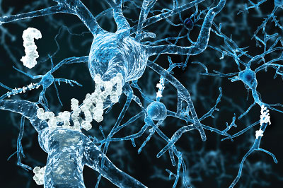
Abnormally folded proteins may contribute to some cases of schizophrenia, according to a study published May 6 in AJP in Advance. This discovery adds to the evidence that schizophrenia is a heterogenous condition with multiple manifestations. The findings also point to a possible relationship between schizophrenia and neurodegenerative disorders; diseases such as Alzheimer’s and Huntington’s also involve abnormally folded proteins.

“When Emil Kraepelin first studied schizophrenia over a century ago, he referred to it as dementia praecox [premature dementia],” said lead investigator Frederick Nucifora, D.O., Ph.D., an assistant professor of psychiatry at Johns Hopkins School of Medicine. “It is interesting to speculate whether he was seeing patients with misfolded, dysfunctional proteins in the brain.”
These findings come from an analysis of postmortem brain tissue from 83 people (42 with schizophrenia and 41 without) collected from three separate brain banks (Harvard Medical School, University of Pittsburgh, and University of Texas Southwestern Medical Center). Nucifora and his colleagues applied techniques used in the study of brain samples from patients with Alzheimer’s to separate brain proteins based on their solubility. Normally, most proteins fold in a way to ensure they stay water soluble; if something goes wrong, misfolded proteins quickly attach to another molecule (usually another misfolded protein) and become inactive. Cells have mechanisms to recycle misfolded proteins, but in some instances, misfolded protein aggregates can overwhelm cells and kill them.
Nucifora got the idea to examine solubility from his clinical experience with a family with a rare, inherited, schizophrenia-like disease. He found that the genetic mutation in this family caused many proteins to misfold and aggregate. “I thought that if misfolding can contribute to this inherited case of schizophrenia, maybe it also happens in sporadic schizophrenia,” he told Psychiatric News. As with Kraepelin, Nucifora had seen many patients with sporadic schizophrenia who also showed a trajectory of cognitive decline that was similar to that experienced by patients with Huntington’s or Parkinson’s.
Nucifora’s hypothesis seemed correct: Almost half of the schizophrenia brain samples (20 of 42) had significantly more insoluble proteins compared with the remaining samples.
The fact that not all the schizophrenia samples had this insoluble-protein problem suggested that antipsychotic medications—which all the patients had taken—were not the cause of this excess insolubility. To further rule out this possibility, the researchers treated rats with haloperidol or risperidone, commonly used first- and second-generation antipsychotics, respectively, for over four months and found that neither drug induced protein insolubility.
The researchers also considered the possibility that the insoluble proteins were an artifact of the postmortem brain preservation process. However, the consistent results seen across samples from all three brain banks, each of which used its own preservation technique, argued against this.
A closer look at the proteins in the insoluble fraction revealed a disproportionate number of proteins involved in the growth and function of axons (the thin fibers that protrude from neurons and transmit electrical nerve impulses to neighboring cells). The abundance of axon-related, insoluble proteins suggests these neurons might have difficulty making proper connections with each other, which is consistent with many theories that schizophrenia results from abnormal brain connectivity.
Of course, postmortem samples can provide only so much information. Nucifora and his team are currently collecting olfactory nerve samples (the only neurons one can readily collect from a living person) from patients with schizophrenia to see whether they can detect insolubility in living patients. If so, his team will compare the clinical profiles of patients with or without insoluble proteins to see whether there are differences in which symptoms are present or how these patients respond to medications.
This study was supported by the Brain and Behavior Research Foundation. ■
“Increased Protein Insolubility in Brains From a Subset of Patients With Schizophrenia” can be accessed here.


