Protein in Blood Offers Clues About TBI Severity
Abstract
Blood concentrations of the neurofilament light protein might help track the progression of recovery following a head injury in athletes and other individuals.

Even if an overdose is accidental, follow-up care is necessary to ensure it does not happen again, says Austin S. Kilaru, M.D., M.S.H.P.
A protein known as neurofilament light chain appears to be a reliable blood-based biomarker for identifying a single traumatic brain injury (TBI) up to years after the injury occurred. This discovery was based on a comprehensive analysis of blood samples from over 300 adults in two separate participant groups. The findings were published in a pair of studies in Neurology in July.
There are currently no proven blood tests that can provide an objective diagnosis of mild TBI or prognosticate the extent of neurological damage from a brain injury. A TBI blood test involving two proteins called UCH-L1 and GFAP was approved by the Food and Drug Administration in 2018, but that test only assesses the risk of intracranial bleeding following a TBI.
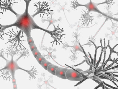
Neurofilament light chain, or NfL, is a structural protein that helps neurons form the long and slender fibers (axons) used to transmit electrical signals across the body. When neurons break down, NfL is released into the environment surrounding them. Research has shown that NfL concentrations in the spinal fluid are excellent proxies for nerve degeneration in the brain, and now many investigators are exploring NfL-based blood tests for diseases such as Huntington’s, multiple sclerosis, and Alzheimer’s.
Pashtun Shahim, M.D., Ph.D., an investigator at the National Institutes of Health Intramural Research Program, wanted to know if measuring NfL levels in blood might also be able to predict physical nerve damage sustained during a concussion. He and colleagues conducted a series of tests in both Swedish athletes and a patient cohort at the NIH Clinical Center.
The first group of participants included 45 Swedish hockey players who had experienced their first concussion in the past week, 31 hockey players with post-concussive syndrome due to multiple head hits, 28 hockey players with no history of concussion, and 14 non-athletes with no history of concussion. All the participants donated blood and spinal fluid samples to measure NfL concentrations.
The analysis showed that NfL levels in both blood and spinal fluid were elevated in individuals who had experienced a TBI compared with controls. Though the NfL levels could not distinguish hockey players who received one versus multiple head injuries, the tests could distinguish players with postconcussive syndrome from concussion-free controls with about 97% accuracy.
Additional analysis on the 45 hockey players who had experienced a concussion the week prior demonstrated that the NfL blood test could distinguish with about 82% accuracy the players whose symptoms resolved within days versus those who had to sit out for an extended period because of prolonged symptoms. The authors noted this could be a clinically important finding as there is an urgent need to find objective tests that can help determine when it is safe for people with concussions to return to work or sports.
The investigators then made use of blood samples from the NIH Clinical Center cohort to assess the long-term and temporal patterns in NfL concentrations. This cohort included 162 individuals with either mild, moderate, or severe TBI as well as 68 TBI-free controls. The participants donated blood samples and received MRI scans at periodic intervals over a five-year period (the first assessment was done 30 days after enrolling in the study).
“This is one of the great aspects of the NIH Clinical Center and a reason I moved from Sweden to continue this research,” said Shahim, who has recently joined the lab of Leighton Chan, M.D., M.P.H., chief of the Rehabilitation Medicine Department at the NIH Clinical Center. “It would be difficult for many academic centers to enroll and maintain such a large patient population for five years.”
The NIH data supported the findings of the Swedish study; average blood levels of NfL rose with the severity of TBI and the blood tests were able to distinguish individuals who had experienced a TBI from controls, even at the five-year assessment. The blood test was most accurate at the 30-day and 90-day assessments, however. At 30 days, the NfL blood test could distinguish controls from people with mild, moderate, or severe TBI with accuracies of 84%, 92%, and 92%, respectively.
For the NIH analysis, the investigators also compared the diagnostic capability of NfL with other potential blood-based biomarkers, namely UCH-L1, GFAP, and the Alzheimer’s protein tau. Of the four proteins, NfL had the highest accuracy in distinguishing participants with TBI from controls and was the only protein to show diagnostic accuracy across all the time points.
Shahim said the findings were quite encouraging but cautioned that there are hurdles and limitations to overcome. Currently, NfL tests would not be useful as a point-of-care test to assess TBI severity in someone admitted to a hospital. “We haven’t analyzed enough people yet to establish a good cutoff between normal and abnormal NfL levels in a given individual,” he said.
NfL blood tests could be valuable, however, in tracking specific individuals over time. For instance, rookie hockey players could provide blood samples at the start of their career that could be used as a point of reference for blood samples collected directly after an injury to see if there are noticeable changes that would signal underlying damage.
The studies were funded by the NIH Intramural Research Program and the Department of Defense’s Center for Neuroscience and Regenerative Medicine. ■


